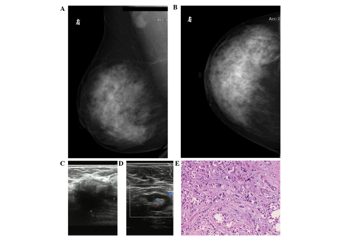
Figure 1.
(A) Oblique and (B) axial mammography findings showing a twisted local structure with sand-like calcification in the superior external quadrant of the right breast. A significantly enlarged lymph node was observed in the right axilla. (C) Echography imaging showing a low echo 2.6×1.8-cm mass with an ill-defined margin. The mass was less regular in morphology, rough and presented with an uneven echo. Color Doppler flow imaging (CDFI) showing no blood flow signal detected in and around the mass. (D) Echography imaging showing a 2.6×1.2-cm hypoechoic nodule found in the right axilla, with a clear edge and a center of echo enhancement, and CDFI showing a small amount of blood flow in the nodule. (E) Lipid-rich carcinoma of the breast, with typical large, vacuolated cells arranged in clusters (hematoxylin and eosin staining; magnification, ×100).

