Figure 8.
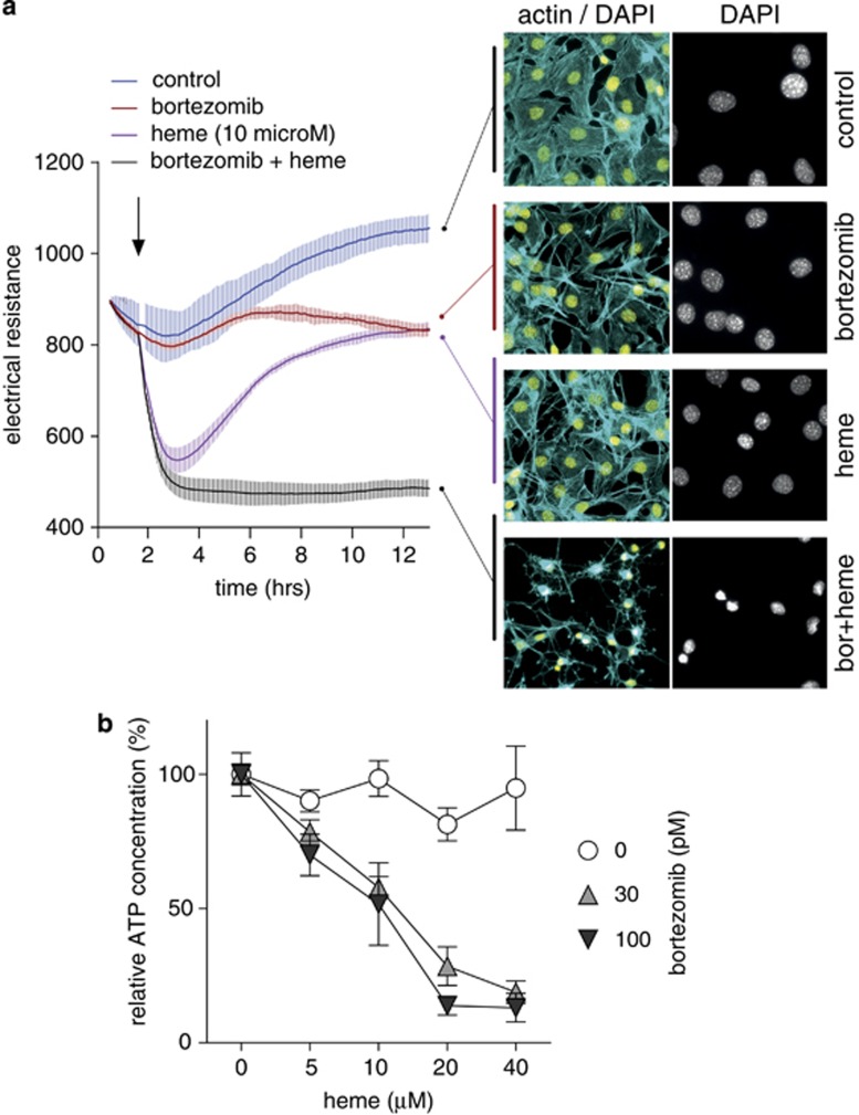
Proteasome inhibition sensitizes cells for heme toxicity. (a) Monolayer resistance of confluent Hmox1 (+/+) MEF cells was measured with an ECIS instrument during incubation of the cells with or without heme (10 μM), bortezomib (100 pM), or the combination of heme+bortezomib. Data represent mean±S.D. of four biologic replicates. At the end of the experiment, cells were fixed and stained for fluorescence microscopy with DAPI (yellow: nuclei) and Alexa 488 phalloidin (cyan: actin cytoskeleton). (b) Hmox1 (+/+) MEF cells were treated with a range of heme concentrations in the presence or absence of bortezomib (30 pM and 100 pM). After 12 h, cellular ATP was measured with a luminescence assay. Data are normalized to the respective control and represent mean±S.D. of six biologic replicates

