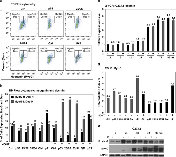
Figure 5.
Repression of myogenin by p53 leads to reduction of late but not early differentiation markers. (a) In response to ectopic p53, expression of an early differentiation marker, desmin, was quantitatively analyzed by co-immunostaining myogenin in RD cells and followed by flow cytometry analysis at 48 h of 4OHT induction. Two gates were drawn as myogenin-low and desmin-high (MyoG-L Des-H) on the left and myogenin-high and desmin-high (MyoG-H Des-H) on the right. (b) Summary of the percentage of RD cells in either MyoG-H Des-H or MyoG-L Des-H. QM: p5325265354. (c) Q-PCR analysis of desmin expression from 2 to 96 h post IR in C2C12 myoblasts maintained under differentiation condition. (d) Expression of MyHC in RD cells in response to ectopic p53 and mutants was analyzed by immunofluorescence (IF) and quantified by the Differentiation Index, which is the percentage of MyHC-positive cells above the total number of nuclei. Ectopic p21 served as a positive control. (e) Immunoblotting shows expression of myogenin and MyHC from 6 to 96 h post IR in C2C12 myoblasts maintained under differentiation condition

