Figure 6.
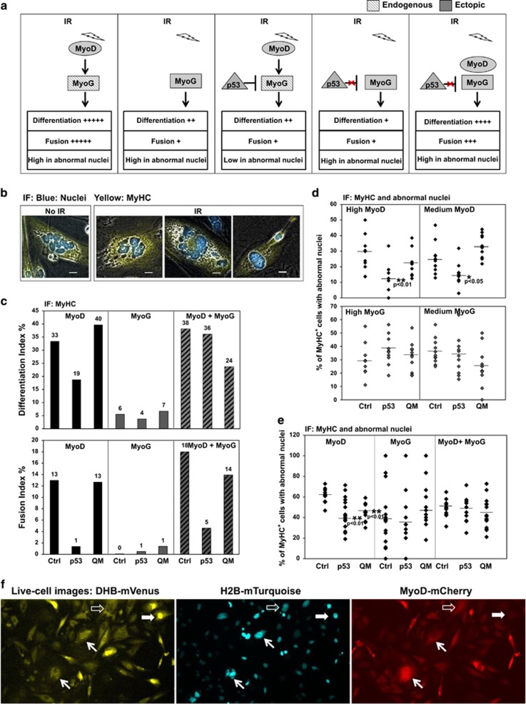
p53-mediated repression of myogenin reduces post-mitotic nuclear abnormality in terminally differentiated cells in response to acute DNA damage. (a) Experimental design and summary of results on p53-null MEFs for assays of myogenin function in either a p53-dependent or p53-independent manner. IR: ionizing radiation. Red-cross sign: p53 is unable to repress ectopic myogenin. Endogenous expression is shown as hashed grey and ectopic expression as filled grey. Myogenic differentiation was quantified using both Differentiation Index (the percentage of MyHC+ cells above the total number of nuclei) and Fusion Index (the percentage of the number of nuclei in MyHC+ cells containing at least three nuclei above the total number of nuclei). The percentage of abnormal nuclei in differentiated muscle cells (MyHC+) is summarized as either high or low in abnormal nuclei. (b) Examples of nuclear abnormality observed in irradiated cells versus non-irradiated cells. Using immunofluorescence (IF), nuclei were scored as abnormal if they were micronuclei, macronuclei, lobulated nuclei, or fragmented nuclei. Scale bar: 20 μm. (c) Myogenic differentiation induced by MyoD, myogenin, or both in the presence of ectopic p53 or the QM mutant, p5325265354. (d) The percentage of abnormal nuclei in MyHC+ cells expressing a high or medium level of MyoD or myogenin in the presence of ectopic p53 or the QM mutant. Statistical significance of reduction in numbers of abnormal nuclei relative to that of the control was determined by a two-tailed Student's t-test. (e) The percentage of abnormal nuclei in MyHC+ cells induced by MyoD, myogenin, or both in the presence of ectopic p53 or the QM mutant. (f) Live-cell images show cells undergoing myogenic conversion and acquiring post-mitotic nuclear defects. Images are shown in three channels from a single frame at 36 h post IR. DHB-mVenus: a fragment of DHB fused to a yellow fluorescent protein. H2B-mTurquoise: a histone H2B fused to a cyan fluorescent protein. MyoD-mCherry: MyoD fused to a red fluorescent protein. Solid white block arrows indicate a cell in G1 and open white block arrows indicate a cell in G2. White slender arrows indicate two MyoD-positive cells showing severely fragmented nuclei arrested in G1 phase at 36 h post IR after one or two rounds of mitosis

