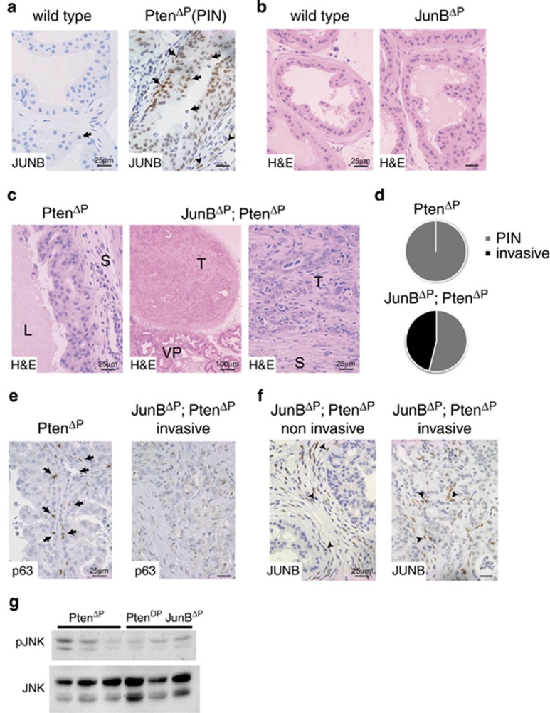
Figure 2.
Loss of JUNB expression induces invasive prostate cancer. (a) IHC for JUNB in normal mouse dorsolateral prostate (wild type) and high-grade PIN (PtenΔP: Ptenflox/flox; PsaCre+/T). Black arrows mark JUNB-positive cells in the epithelia. n≥3. A representative sample is shown. (b) Haematoxylin and eosin (H&E) staining of dorsolateral prostate from wild-type, JunBΔP (JunBflox/flox; PsaCre+/T) mice; n≥3. A representative sample is shown. (c) H&E staining of dorsolateral prostate from PtenΔP (Ptenflox/flox; PsaCre+/T) and JunBΔP; PtenΔP (Junbflox/flox; Ptenflox/flox; PsaCre+/T) mice. L (lumen), T (tumour), S (stroma) and VP (ventral prostate). n≥3. A representative sample is shown. (d) Incidence of invasive prostate cancer in 12-week-old mice. n= PtenΔP: 0 out of 12; JunBΔP; PtenΔP: 6 out of 13. (e) Staining for basal cells with an antibody to p63 (brown). A representative noninvasive sample and an invasive sample are shown; n=5. (f) IHC for JUNB in noninvasive and invasive prostate cancer from JunBΔP; PtenΔP (Junbflox/flox; Ptenflox/flox; PsaCre+/T) mice. Black arrowheads mark JUNB-positive cells in the stroma. n≥3. A representative sample is shown. (g) Western blot for phospho-JNK in prostate samples from PtenΔP and JunBΔP; PtenΔP mice. Total JNK is used as loading control; n=3

