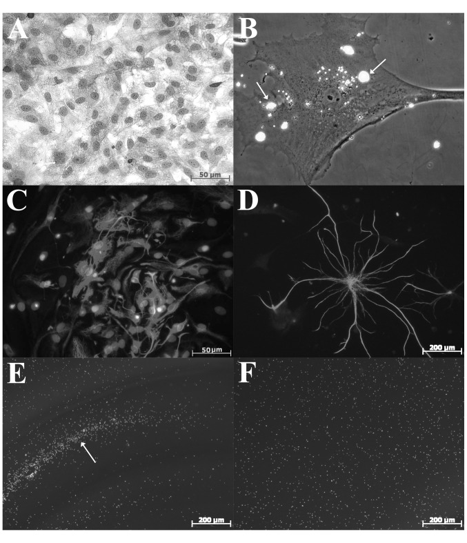
Figure 3.
Characteristics of cell lines and control fibroblast cultures and astrocytes, and reaction of different cell lines to co-culturing with glioma cells in the experiment. (A) Rat fibroblasts (hematoxylin and eosin; magnification, ×40). (B) Rat fibroblasts. Phagocytosis of collagen immobilized on the surface of the fluorescent microparticles FluoSpheres® Collagen I-Labeled Microspheres (magnification, ×630). (C) Rat astrocytes stained with anti-GFAP Mab and nuclei counterstained with DAPI (magnification, ×100). (D) Rat astrocyte stained with anti-GFAP Mab (magnification, ×200). (E) Formation of fluorescent cell shaft on the perimeter of culture inserts containing glioma cells, by hematopoietic stem cells stained with Vybrant® CFDA SE Cell Tracer (magnification, ×10). (F) Co-culturing hematopoietic stem cells and rat fibroblasts. The formation of the cell shaft was absent (magnification, ×10). GFAP, glial fibrillary acidic protein; Mab, monoclonal antibody.

