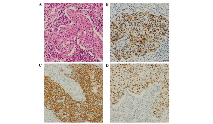
Figure 2.
Histological features of lymphoepithelioma-like carcinoma of the lung. (A) Histological examination revealed a large island of tumor cells infiltrated by intense lymphoplasmacytic cell population (HE; magnification, ×400). (B) Cells were positive for Epstein-Barr virus (EBV)-encoded small nonpolyadenylated RNA (magnification, ×400). Immunohistochemical staining revealed that the specimen cells were positive for (C) cytokeratin 5/6 (magnification, 400x) and (D) P63 (magnification, ×400). HE, hematoxylin and eosin.

