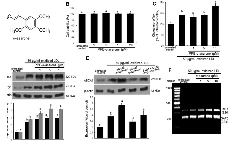
Figure 5.
(A) Chemical structure, (B) cytotoxicity of α-asarone, (C) enhancement of cholesterol efflux by α-asarone, (D and E) upregulation of ABCA1 and ABCG1 by α-asarone and β-asarone, and (F) elevation of retinoid X receptor (RXR)α transcription. J774A.1 murine macrophages were exposed to 50 μg/ml oxidized low-density lipoprotein (LDL) and treated with 1–10 μM purple Perilla frutescens extracts (PPE)-α-asarone and 5–10 μM β-asarone. (B) MTT assay was performed for the measurement of α-asarone toxicity. Graph data represent 1 of 4 independent experiments with multiple estimations. Values are expressed as the percentage cell survival relative to the untreated control cells (cell viability, 100%). (C) Cholesterol efflux was expressed as the percentage fluorescence in the medium relative to total fluorescence. (D and E) For the measurement of ABCA1 and ABCG1 expression, total cell lysates were subjected to western blot analysis with a primary antibody against ABCG1 or ABCG1. β-actin was used as an internal control. Bar graphs (means ± SEM, n=3) represent quantitative densitometric results of the upper bands. Bar graphs denoted without a common letter indicate significant difference, P<0.05. (F) RXRα mRNA expression was measured by RT-PCR. GAPDH was used as a housekeeping gene for the co-amplification with RXRα.

