
Figure 3. Retinal phenotypes of children with PNPLA6 mutations.
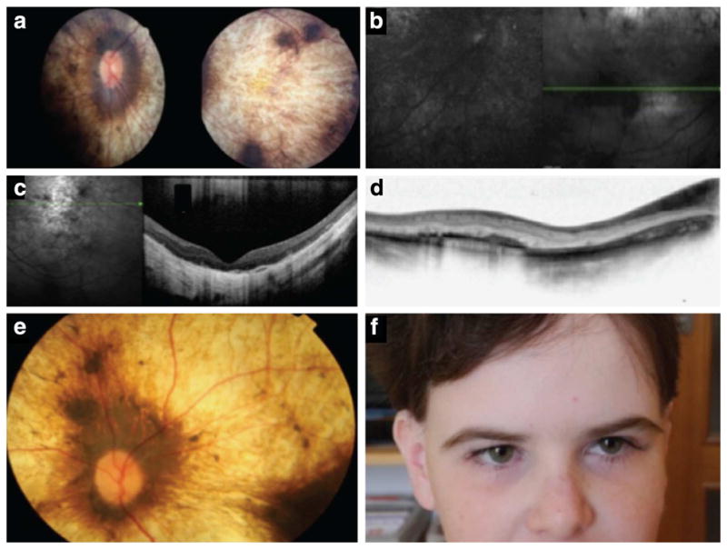
(a) A retinal photo shows severe choroidal and retinal atrophy ‘choroideremia like’ in patient 5,267. (b) Fundus autofluorescence (FAF) shows severely abnormal and grossly absent lipofuscin metabolism. (c) Optical coherence tomography reveals severe photoreceptor loss, retinal thinning and retinal remodelling. (d) Using optical coherence tomography, we found that the inner retinal layers are also abnormal. (e) Another child (167) with mutations in PNPLA6 shows a strikingly similar retinal appearance as shown in a, again illustrating the ‘choroideremia-like’ retinal changes. (f) Full-face photo of a child with PNPLA6 mutations, illustrating the extremely long lashes.