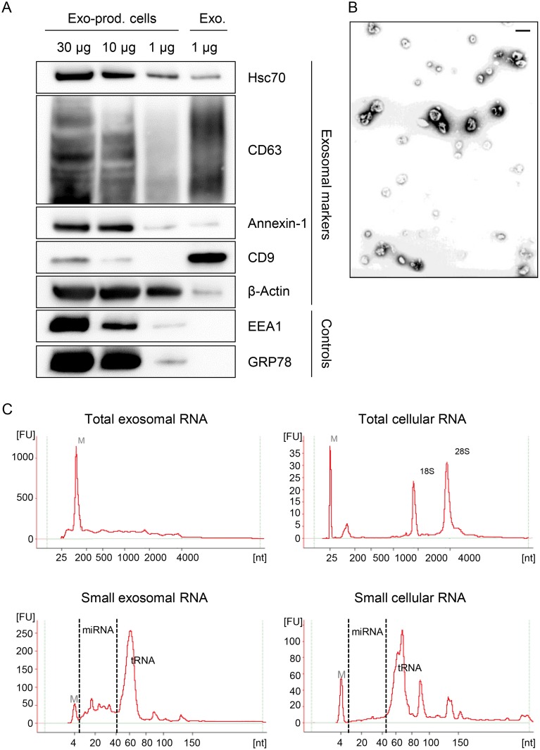
Fig 7. Characterization of exosomes secreted by HeLa cells used for small RNA deep sequencing.
(A) Immunoblot analysis of total cellular extract (30, 10 and 1 μg) from exosome-producing cells, and of 1 μg protein from exosome preparations. Hsc70, CD63, Annexin-1, CD9 and β-Actin: exosomal markers; EEA1: early endosome marker; GRP78: ER marker. (B) Visualization of exosomes by electron microscopy. Bar corresponds to 100 nm. (C) Characterization of cellular and exosomal RNA. Electropherograms of total RNA isolated from HeLa cells and from RNAse A-treated exosomes. Upper panel: total RNA contents; lower panel: small RNA contents. M = marker. Shown are representative images for siContr-1-treated samples.

