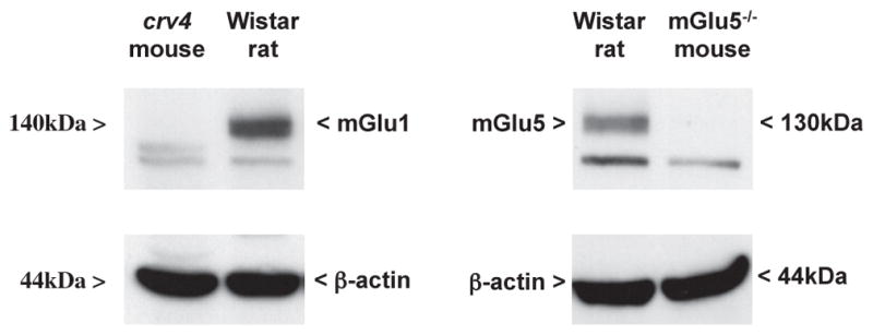
Fig. 5.

Specificity of the antibodies used for immunoblot analysis of mGlu1α and mGlu5 receptors. Western blot analysis was performed in protein extracts from the cerebellum of crv4 mutant mice (left) and the cerebral cortex of mGlu5−/− mice (right) to verify the identity of the bands corresponding to mGlu1α and mGlu5 receptors, respectively. mGlu1α and mGlu5 receptor labelling are also shown in the cortex and thalamus of Wistar rats, respectively, for comparison. The upper band at 140 and 130 kDa corresponds to mGlu1α and mGlu5 receptor monomers, respectively. The lower band was non-specific because it was still present in crv4 and mGlu5−/− mice.
