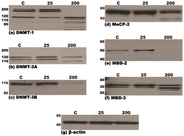
Fig. 2.
Immunoblots demonstrating steady-state levels of DNMT-1 (a), DNMT-3a (b), DNMT-3b (c), MeCP-2 (d), MBD-2 (e) and MBD-3 (f) proteins in nuclear extracts derived from murine embryonic fibroblasts following treatment (48 h) with either 25 mM or 200 mM ethanol or vehicle control (PBS). Equal amounts of protein (15 μg) were resolved by SDS-PAGE on 12% polyacrylamide bis–tris gels, transferred to PVDF membranes, probed with specific antibodies and immunoreactive species detected by chemiluminescence, as detailed in Section 2. Molecular weights of the marker proteins are indicated to the left of each panel. The lowermost panel (g) depicts one representative immunoblot of the normalization control, β-actin. Each immunoblot is representative of no less than three independent blots from three unique sets of extracts from control and ethanol treated MEF cells.

