Figure 1.
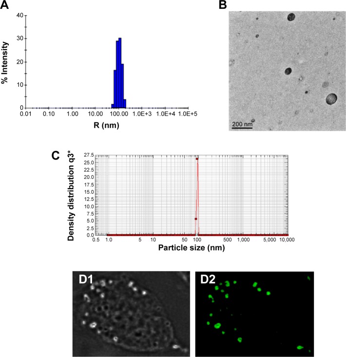
Size distribution and morphology of AmB–PGA nanoparticles.
Notes: (A) The Z-average diameter of the AmB–PGA nanoparticles determined by dynamic light scattering measurements was 107±0.7 nm. (B) Transmission electron microscopy showed that the AmB–PGA nanoparticles were spherical in shape with an average size of 98±2 nm. (C) Particle size distribution of AmB–PGA nanoparticles assessed by photon correlation spectroscopy. (D1) Phase contrast light and (D2) fluorescence micrographs of normal peritoneal macrophages after their interaction with calcein (fluorescent probe)-loaded PGA nanoparticles.
Abbreviations: AmB, amphotericin B; PGA, polyglutamic acid.

