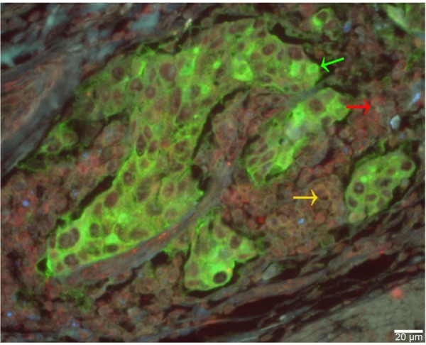
Figure 2.

Multicolor QD-based IHC staining for GC tissue.
Notes: GC tissue marked with cancer cell are green (green arrow), with macrophages are yellow (yellow arrow), and with lymphocytes are red (red arrow). Scale bar: 20 μm. Magnification for all images: 400×.
Abbreviations: QD, quantum dot; IHC, immunohistochemistry; GC, gastric cancer.
