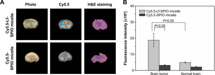
Figure 7.

Ex vivo study of the brain tissue.
Notes: (A) Ex vivo fluorescence images and H&E-staining images of rat brain bearing C6 glioma at 48 hours postinjection. (B) The average fluorescence intensity of the brain tumor and the normal brain. Results expressed as means ± standard error of mean (n=6).
Abbreviations: Lf, lactoferrin; SPIO, superparamagnetic iron oxide; H&E, hematoxylin and eosin.
