Figure 3.
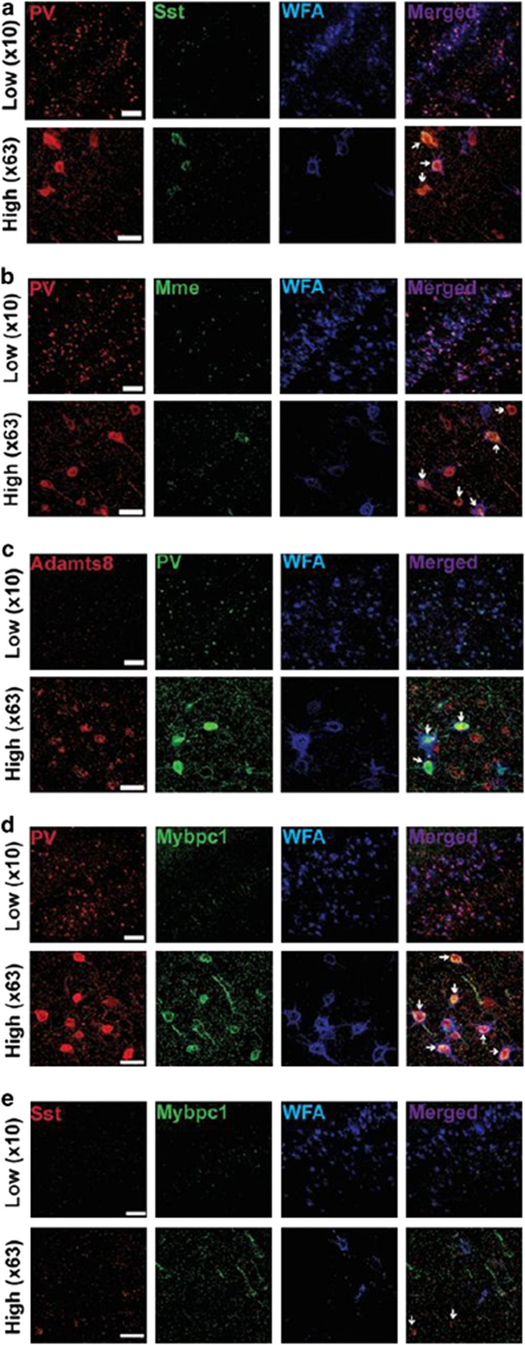
Immunofluorescence and confocal imaging. Parvalbumin (PV) interneurons enwrapped with perineuronal net contain metallopeptidase protein. Colocalization of PV, somatostatin (Sst), Wisteria floribunda agglutinin (WFA), Mme (Neprilysin), Adamts8 and Mybpc1. (a) Micrograph of parvalbumin interneurons (red) revealing that most PV coexpressed with Sst (green) are void of perineuronal net (WFA; blue). (b) Parvalbumin interneurons (red) enwrapped with perineuronal net (blue) contain Neprilysin (Mme; green). (c) Adamts8 (red) can be found in all parvalbumin interneurons (green), but not exclusively. (d) The majority of parvalbumin interneurons (red) enwrapped by perineuronal net (blue) colocalize with Mybpc1 (green). (e) Somatostatin-positive neurons (red) are not enwrapped by perineuronal net (WFA, blue) and do not colocalize with Mybpc1 (green). Scale bar, 80 μm in low ( × 10) and 30 μm in high ( × 63) magnification. Arrows depicts main cell of interests (red). This observation was confirmed in three male 40-day-old C57/Bl6 mice.

