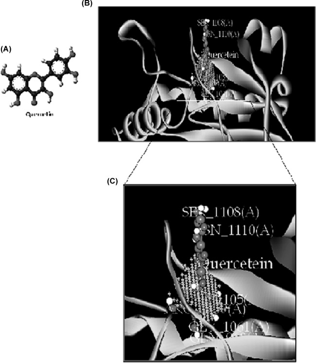
FIG 9.
Molecular docking of the interaction between QC and adenylate cyclase. (A) 3D structure of QC. (B) Binding orientation of QC in adenylate cyclase. The protein is depicted as a ribbon, and secondary structures (i.e., helix, strand, and loop) are shown. (C) Both QC and ligand contact residues are represented in stick form.

