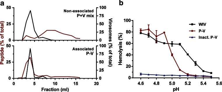
Fig. 2.
Characteristics of peptide-loaded virosomes. Peptide association of peptide mixed with virosomes (P+V mix) and peptide-loaded virosomes (P-V) analyzed by size exclusion chromatography (a). Black lines show the virosome elution pattern (based on protein determination), whereas the red line shows the elution of peptide (based on fluorescence of M158–66-FITC). The fusogenic activity of WIV (black), P-V (red) and fusion-inactivated P-V (blue) was determined between pH 4.6 and 5.5 by a hemolysis assay (b). Data represents mean ± SD (n = 3).

