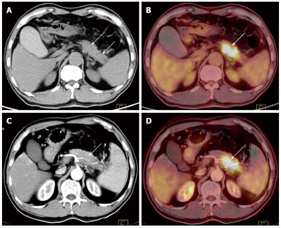
Figure 2.

A 61-old male patient with upper abdominal pain for more than 3 mo. A-B: PET/CT image [A: Enlargement at the junction of the pancreatic body and tail (depicted by plain CT scanning); B: Increased FDG uptake at the lesion with the SUVmax of 10.6. This disease was diagnosed as pancreatic cancer (depicted by a PET/CT image)]; C-D: CECT and PET/CECT fusion image: splenic artery invasion was clearly displayed; the splenic artery was thinner with irregular vascular edges. This case was pathologically diagnosed as a moderately differentiated pancreatic ductal adenocarcinoma with splenic artery invasion. PET/CT: Positron emission tomography/computed tomography; CECT: Contrast-enhanced CT; FDG: Fluorodeoxyglucose.
