Figure 3.

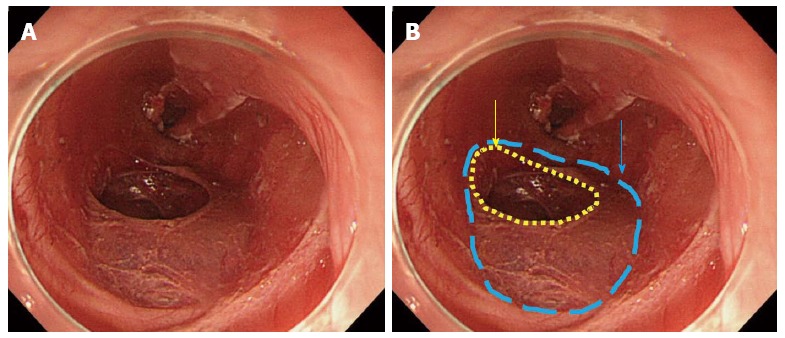
After removing dissected tissue. A: Ulcer bed after endoscopic submucosal dissection; B: Blue dotted line indicates the defect of the muscularis propria layer, and yellow dotted line indicates the perforation hole.

After removing dissected tissue. A: Ulcer bed after endoscopic submucosal dissection; B: Blue dotted line indicates the defect of the muscularis propria layer, and yellow dotted line indicates the perforation hole.