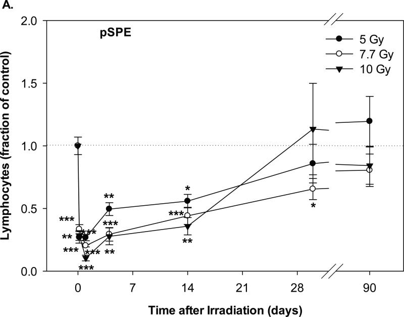
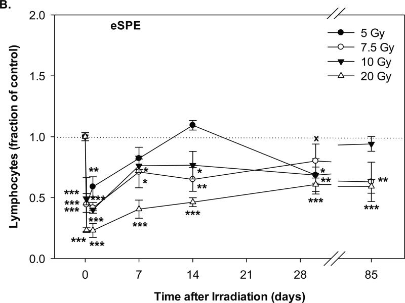
Fig. 2.
Change of lymphocyte count in Yucatan minipigs exposed to pSPE radiation or eSPE radiation. A minimum of 3 animals per group were exposed to pSPE radiation (A) at a single skin dose of 5 , 7.7 or 10 Gy or eSPE radiation (B) at a single skin dose of 5, 7.5, 10, or 20 Gy. The lymphocyte count was determined at the indicated time points. The results are expressed as fraction of control and compared with the pre-irradiation baseline value (indicated by dashed line). Statistical significance of the comparison is indicated by an x (p is between 0.005 and 0.10, and considered as marginally significant), * (p < 0.05), ** (p < 0.01) and *** (p < 0.001), as appropriate. Each data point represents a mean of three animals, and the associated standard error is indicated by the error bar.


