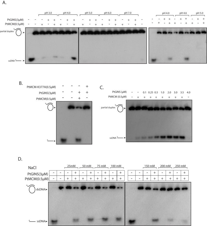
Figure 5. Helicase assays.
(A). Reactions carried out with 500 nM PtMCM in presence and absence of PtGINS (3000 nM) in 30 mM Tris-acetate buffers of different pH. Reactions were analyzed on three separate 6% gels, and run under identical conditions of voltage. Cropping lines indicate separate gels. Lane 1 of first and last gels - radiolabelled ssDNA only (loaded as marker); lane 2 of first gel and last lane of second gel– helicase reactions incubated in absence of protein (B). Reactions carried out with 500 nM PtMCM (wild type and K337A mutant) and 3000 nM PtGINS in 30 mM Tris-acetate buffer of pH 4. Lane 1- radiolabelled ssDNA only (loaded as marker); lane 2 – helicase reaction incubated in absence of protein (C). Reactions carried out with 500 nM MCM and increasing concentrations of GINS in 30 mM Tris-acetate buffer of pH 4. Lane 1 – helicase reaction incubated in absence of protein (D). Reactions carried out with 500 nM PtMCM in presence and absence of 3000 nM GINS in 30 mM Tris-acetate buffer of pH 4; NaCl concentrations in the reaction varied from 25 mM to 250 mM. Reactions were analyzed on two separate 6% gels, and run under identical conditions of voltage. Cropping lines indicate separate gels. Lanes 1 of both gels- radiolabelled ssDNA only (loaded as marker); lanes 2 of both gels – helicase reaction incubated in absence of protein. All reactions were performed at 55°C for 1 h. Composition of helicase assay buffers detailed in Methods.

