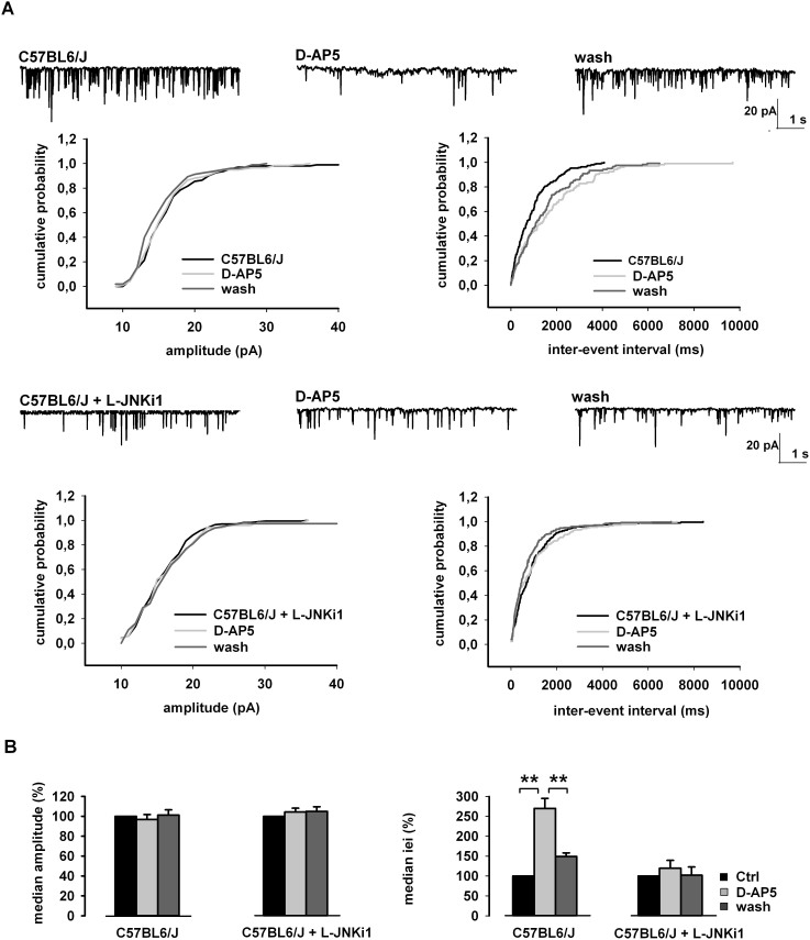
Figure 4. Functional role of JNK in NMDA-dependent glutamate release.
(A) Cumulative distributions of mEPSC amplitude (left) and inter-event interval (iei) (right) recorded from a single C57BL6/J neuron, or C57BL6/J neuron pre-incubated with L-JNKi1 in response to D-AP5. The traces on top are obtained from the same neuron, in control conditions, during D-AP5 application and at D-AP5 washout. (B) Histograms of the median values expressed as percentage versus control (100%) of mEPSC amplitude (left) and iei (right) recorded from 11 neurons of C57BL6/J, and 10 neurons of C57BL6/J pre-incubated with L-JNKi1 (n = 10) in response to D-AP5. **p < 0.01 (t-test).

