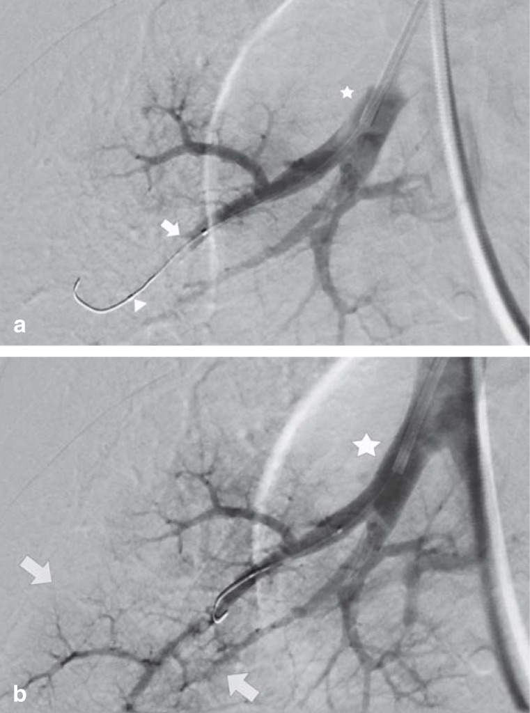
Figure 4.
Pulmonary balloon angioplasty of the A8 segment of the right lower lobe.
a) Selective imaging of the right A8 segmental artery, in which the contrast is visibly interrupted at the right cardiac margin (arrow). The stenosis has already been successfully passed with a wire (arrowhead).
b) Selective DSA series (DSA, digital subtraction angiography) to follow up treatment outcome after dilatation of web stenosis with a suitable balloon catheter. There is clear contrast of the peripheral branches of the A8 segmental artery, with visualization of even the smallest subbranches of the pulmonary artery (arrows). The location of the guide catheter is the same in both series (*)

