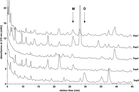
Figure 4.

HPLC profiles of the fragmentation of LK by selected Saps. LK samples (1.5 μM) were digested with 0.03 μM of Sap1, −3, −4, −8, and −9 in the citrate buffer (pH 5.0) at 37°C for 24 hours. The reaction was stopped using pepstatin A (10 μM), followed by acidification with HCl (0.33 M). The samples were analyzed using HPLC on the Luna C18(2) 5 μm 4.6 × 250 mm column (Phenomenex) in a TFA water-ACN binary gradient system, as described in the Materials and Methods section. Arrows indicate the retention times of the Met-Lys-bradykinin (M) and des-Arg1-bradykinin (D) standards.
