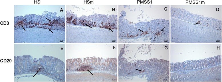
Figure 4.

Gastric inflammation of Helicobacter -infected Mongolian gerbils. CD3 staining of the antrum of the stomach from Mongolian gerbils inoculated with H. suis HS5cLP (A), H. suis HS5cLPΔggt (B), H. pylori PMSS1 (C) and H. pylori PMSS1Δggt (D) at 9 weeks post inoculation, showing T-lymphocytes (brown). CD20 staining of the antrum of a gerbil infected with WT H. suis HS5cLP (E), H. suis HS5cLPΔggt (F), WT H. pylori PMSS1 (G) and H. pylori PMSS1Δggt (H) at 9 weeks post inoculation, showing B lymphocytes (brown) in germinal centers of lymphoid follicles (arrows) or lymphoid aggregates (arrows). HS: animals infected with WT H. suis strain HS5cLP; HSm: animals infected with H. suis strain HS5cLPΔggt; PMSS1: animals infected with WT H. pylori PMSS1; PMSS1m: animals infected with H. pylori PMSS1Δggt; WT: wild-type. Original magnification: 100 × .
