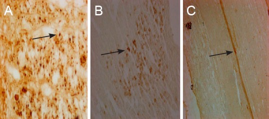
Figure 7.

Neuronal retrograde tracing and nerve fibers at the spinal cord hemisection injury site after combined biological conduit (biotin dextran amine staining, light microscope).
(A) Morphology of normal cerebral cortex (× 400); (B) morphology of cerebral cortex in the biological chitin tube group 14 weeks after surgery (× 200), showing biotin dextran amine-positive fibers, terminals and cells in the contralateral side to the injection site; (C) strongly labeled spinal cord nerve fibers in the biological chitin tube group 14 weeks after surgery against a pale yellow background, showing a clear distinction between labeled fibers and terminals (× 100). Arrows refer to biotin dextran amine-positive cells.
