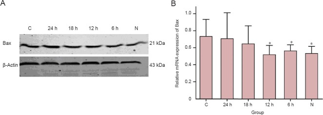
Figure 3.

Mild hypothermia decreased pro-apoptotic protein Bax expression in neurons subjected to oxygen-glucose deprivation/reperfusion.
(A) Western blot analysis demonstrating expression patterns of indicated protein bax in the lysate of each cell group, with β-actin used as a loading control. (B) Data presented in the graph. The values were normalized with β-actin. *P < 0.05, vs. C group. Each experiment was repeated three times, and one-way analysis of variance followed by the least significant difference was used for difference comparison between each group. 24, 18, 12, 6 h: 24, 18, 12, 6 hours of hypothermia treatment; N: normal; C: control.
