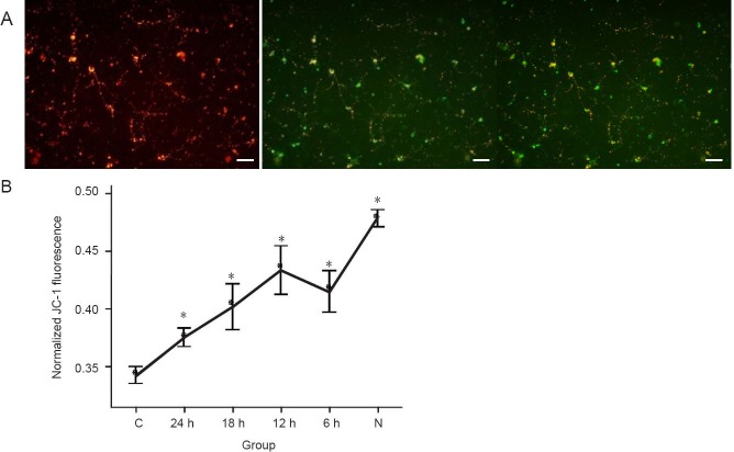
Figure 4.
Mild hypothermia increased mitochondrial membrane potential (MMP) of neurons subjected to oxygen-glucose deprivation.
(A) If MMP depolarizes, JC-1 (Reers et al. (1991), a useful tool used for MMP), becomes a monomer (green), and if it polarizes, it becomes a compound (red). This voltage-sensitive dye follows the Nernst behavior, and increased uptake of this probe is caused by MMP. When illuminated with 490-nm and 525-nm light, JC-1 emits peaks at 530 nm (green) and 590 nm (red) respectively. The ratio between green and red depends on MMP. Scale bars: 50 μm. (B) Sequential change of normalized JC-1 fluorescence after 90 minutes of oxygen-glucose deprivation and reperfusion. The ratio was obtained by comparison with the value of the compound (red)/monomer (green) ratio in the group. *P < 0.05, vs. C group, one-way analysis of variance followed by the least significant difference test was performed to compare the difference between each group. 24, 18, 12, 6 h: 24, 18, 12, 6 hours of hypothermia treatment; N: normal; C: control.

