Figure 1.

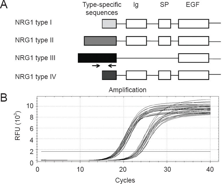
Diagram of neuregulin-1 (NRG1) gene structure and amplification curves of quantitative real-time polymerase chain reaction.
(A) NRG1 alternative splicing variants and domains. Primers designed for detection of NRG1 type III mRNAs are also shown as arrows. Ig: Immunoglobulin-like domain; SP: spacer domain; EGF: epidermal growth factor-like domain. (B) A polymerase chain reaction baseline subtractive curve fit view of the data was shown with relative fluorescence units (RFU) plotted against cycle number. Arrows refer to the position and direction of NRG1 type III specific mRNA primers.
