
Figure 2.
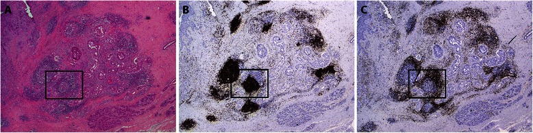
Representative overview of lymphocytic infiltrate in breast tumor. Histology and immunohistochemical analyses performed on breast tumor biopsies show the localization and distribution of lymphocytes in primary tumor of the breast. A) HE staining shows intratumoral TLS with GC, B) CD20+ B lymphocytes forming follicles with surrounding area of C) CD3+ T lymphocytes, resembling highly organized structures of secondary lymphoid tissue. Magnification 20X. Higher magnification of boxed area is shown in Figure 3.