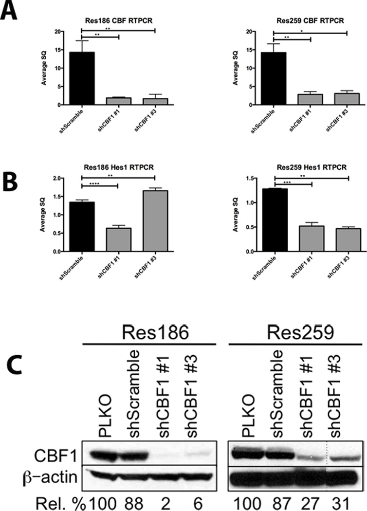
Figure 4.
Notch pathway inhibition through CBF1 knockdown in Res186 and Res259 cell lines. (A) RT-PCR demonstrates a significant decrease in CBF1 mRNA in cell lines Res186 and Res259 using 2 separate short-hairpins. (B) RT-PCR for HES1 mRNA demonstrates a concomitant decrease. (C) Western blot after 7 days of selection shows that CBF1 is also inhibited at the protein level. Representative images of each experiment are shown; each was performed in triplicate after 7 days of selection in 5 µg/ml puromycin. Densitometry analyses representing percent CBF1 knockdown are listed below the images (*p < 0.05, **p < 0.005, ***p < 0.0005, ****p < 0.0001). PLKO, control.

