
Figure 2.
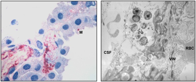
Pathologic findings in the CP of human patients with meningococcal disease. An immunohistochemical examination (left panel) shows abundant bacteria and bacterial antigens in the lumen of a thrombosed blood vessel and in the interstitial tissue. One N. meningitidis organism (M) can be seen in the surface of a CP epithelial cells. Transmission electron microscopy (right panel) demonstrates meningococci (M) in the interstitial space. The blood vessel wall (VW), a red blood cell (RBC) and the space containing cerebrospinal fluid (CSF) are pointed out. The pictures are reproduced with friendly permission from Guarner et al. (2004).