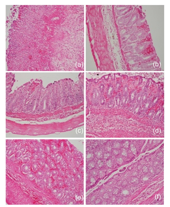
Fig. 3.
Histological sections of rat colon
Rats were treated intrarectally with 4% acetic acid alone (a), or following 100 mg/kg sodium butyrate orally (b), intraperitoneally (c), or intrarectally (d and e). (a) Massive hemorrhage, loss of epithelial lining, loss of crypt and villi, and massive infiltration of inflammatory cells due to acetic acid; (b) Considerable intact epithelium under which there is moderate hemorrhage between crypts and villi, loss of some goblet cells, and moderate infiltration of inflammatory cells in the submucosa; (c) A more intact epithelium, mild hemorrhage, and mild infiltration of inflammatory cells; (d) A considerable intact epithelium with severe hemorrhage underneath and destroyed villi and some crypts, and severe loss of goblet cells in villi, severe cellular infiltration in the submucosa; (e) Intercryptal hemorrhage and retention of crypt architecture; (f) The photomicrographs also include those of control rats treated with sodium butyrate alone with no significant changes. Each photo represents 8 animals (4 animals per treatment in duplicate experiments). Hematoxylin and eosin, original magnification ×200

