
Fig 3. Sublethal caspase activation promotes c-Kit + cardiac progenitors proliferation.
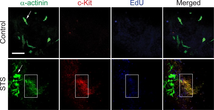
Counterstaining for α-actinin and c-Kit in EBs exposed to 100 nM STS for 5 h and then cultured for additional 7 days. Area defined by the rectangle indicates clusters of c-Kit/α-actininlow cells, and arrow indicates α-actininhigh cardiomyocytes. 7 days after the indicated treatments, EBs were pulsed for 30 minutes with EdU and then stained for α-actinin (green), c-Kit (red) and EdU (blue). Blue nuclei indicate proliferating cells. Bars represent 100 μm.