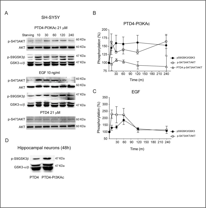
Fig 4. The PI3K activator peptide PTD4-PI3KAc activates AKT and induces phosphorylation of GSK3 at S9.
A. SH-SY5Y cell cultures were serum starved for 16 hours and challenged afterwards with PTD4-PI3KAc 21 μM, EGF 10 ng/mL or PTD4 (control peptide at a concentration of 21 μM). Cells were lysated at the indicated times and membranes incubated with anti-p-S473AKT and anti-p-S9GSK3β. Levels of phosphorylation were normalized to AKT and GSK3β (lower band), respectively. B. Quantitative measurements of p-S473AKT (open circles) and p-S9GSK3β (closed circles) after PTD4-PI3KAc treatment (n = 4). The level of p-S473AKT after PTD4 treatment (open triangles) is included as a negative control (n = 3). C. Quantification of p-S473AKT (open circles) and p-S9GSK3β (closed circles) levels after EGF (10 ng/ml) treatment (n = 5). D. Hippocampal neuronal cultures of 12 DIV were treated for 48 hours with the PTD4-PI3KAc or PTD4 peptides. The treatment results in an average increase of p-S9GSK3β levels of 121 ± 3% (n = 7 from 7 different cultures). Student’s two-tailed t-test, ***p<0.0001.

