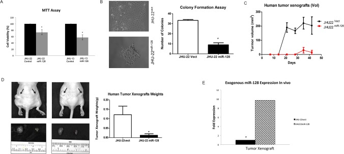
Fig 4. Evaluation of miR-128 effects on cell viability, proliferation and xenograft growth.
(A) The cell viability levels were analyzed by MTT. The results represent the mean ± SD of five independent experiments (*P < 0.05). (B) The cell proliferation levels were determined by a colony formation assay. The colonies were viewed under optic-microscope at day-9 (left) and the colony number was counted (right). (C) The day of cell inoculation was the experimental start day and all mice were sacrificed on day 42. (D) Whole body imaging with tumor xenografts JHU-22vect or JHU-22miR128 cells, 1 x 10⁶ cells in 100 μl, were inoculated subcutaneously into the lower back of the mice. JHU-22vect cells on the left and JHU-22miR-128 cells on the right. The arrows indicated the tumor locations. Tumor mass was measured on the final experimental day immediately after the tumor tissue was removed from the mouse by surgical excision. Results are presented as the mean ± SD, n = 10. (E) Levels of miR-128 in cultured cells and tumor xenografts were determined using QRT-PCR.

