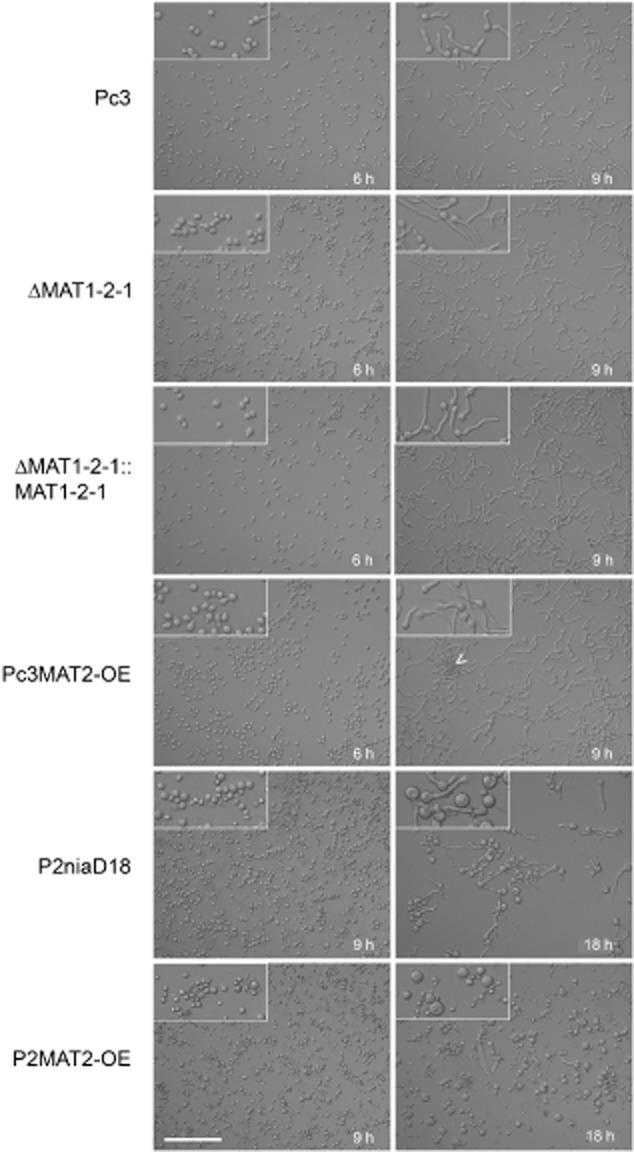
Figure 3.

Micrographs of germinating conidia and formation of spore clusters in strains as indicated. Insets show enlargements (twofold) of images, and the arrowhead points to agglutinated conidiospores. Scale bar corresponds to 100 μm in all images.
