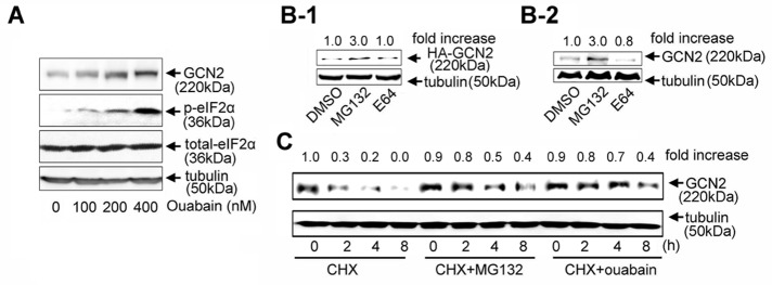
FIGURE 3:

GCN2 ubiquitination and degradation. (A) A549 cells were treated with ouabain for 8 h at 0, 100, 200, and 400 nM. (B) A549 cells transfected (B-1) or nontransfected with HA-GCN2 (B-2) were treated with MG132 (20 μM), E64 (5 μM), or dimethyl sulfoxide for 12 h and the lysates subjected to immunoblot analysis by using antibodies against GCN2 and tubulin. (C) A549 cells were treated with CHX (2 μM) plus vehicle, CHX (2 μM) plus MG132 (20 μM), and CHX (2 μM) plus ouabain (100 nM) for the indicated times and GCN2 protein levels examined by immunoblot analysis. Similar results from three independent experiments are shown.
