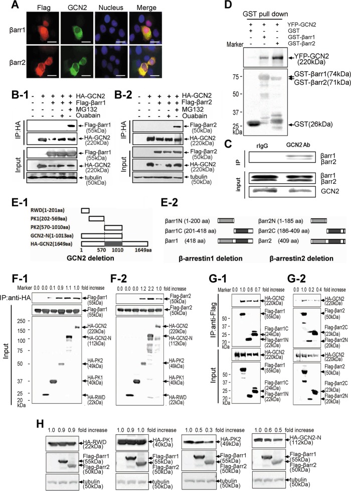
FIGURE 5:
The protein interaction between β-arrestin1/2 and GCN2. (A) A549 cells were cotransfected with YFP-tagged GCN2 and FLAG-tagged β-arrestin1/2. Immunofluorescence analysis was performed as described in Materials and Methods. Scale bar, 20 μm. (B) Cells were cotransfected with expression vectors harboring HA-GCN2 plus FLAG-β-arrestin1 (B-1) or FLAG-β-arrestin2 for 36 h (B-2) and untreated or treated with MG132 (20 μM) or ouabain (200 nM) for 8 h before being harvested. The cell lysates were immunoprecipitated with anti-HA IgG, and the immune pellets were detected by immunoblot analysis with anti-FLAG IgGs. (C) After treatment with 20 μM MG132, A549 cell lysates were used for immunoprecipitation with an anti-GCN2 IgG or normal rabbit IgG and analyzed by immunoblot analysis with anti–β-arrestin1/2 IgGs. (D) YFP-GCN2–transfected HEK293 cells lysates were precipitated with glutathione beads and incubated with purified GST, GST-β-arrestin1, or GST-β-arrestin2. Precipitates were analyzed by immunoblot analysis with an anti-YFP IgG. (E) Deletion mutants for GCN2 (E-1) and β-arrestin1/2 (E-2). (F) HEK293 cells were cotransfected with GCN2 deletion mutants plus FLAG-β-arrestin1 (F-1) or FLAG-β-arrestin2 (F-2). The cells were treated with MG132 (20 μM) for 5 h before being harvested, and lysates were immunoprecipitated with an anti-HA IgG, and the immune pellets were detected by immunoblot analysis using anti-FLAG IgG. (G) A549 cells were cotransfected with HA-GCN2 plus β-arrestin1 deletion mutants (G-1) or β-arrestin2 deletion mutants (G-2). The cells were treated with MG132 (20 μM) for 5 h before being harvested, and lysates were immunoprecipitated with anti-FLAG IgG; the immune pellets were detected by immunoblot analysis with anti-HA IgG. (H) HEK293 cells were cotransfected with HA-RWD, HA-PK1, HA-PK2, or HA-GCN2-N plus FLAG tagged β-arrestin1/2. The lysates were subjected to immunoblot analysis by using the indicated antibodies. A representative Western blot for each treatment from three independent experiments is shown.

