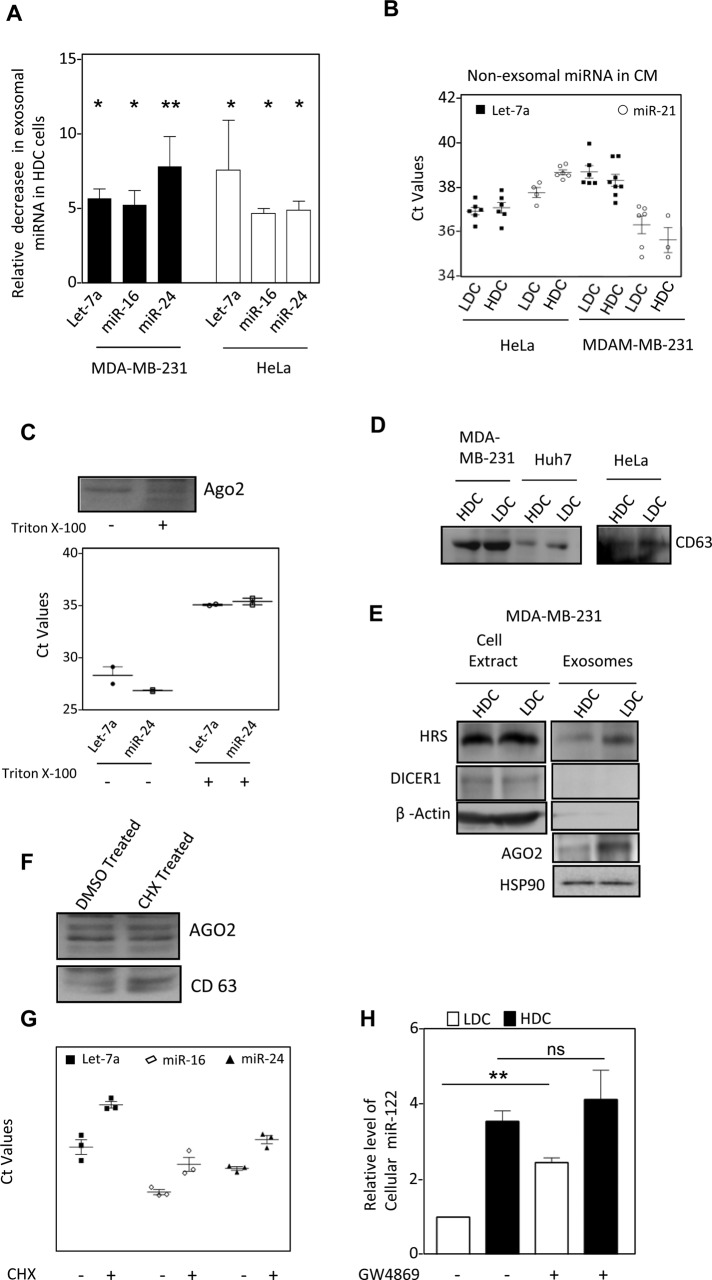
FIGURE 6:
Exosomal export of miRNA is curtailed in cells grown to high density (A) Levels of let-7a, miR-16 and miR-24 miRNAs in FACS sorted identical number of exosomes released by HeLa and MDA-MB-231 cells grown to LDC or HDC cells (mean+/– SEM, n = minimum 3). (B) Amount of miRNA present in cell culture supernatant of HDC and LDC cells after removal of exosomes. (C) miRNA and AGO2 present in cell culture supernatant of HeLa cells is membrane protected. Conditioned medium from LDC state HeLa cells were treated with Triton X-100 for 15 min before they were used for exosome isolation. The estimation of miRNA was done by real time quantification and Ct values are plotted. AGO2 levels were detected by Western blot analysis. (D) Levels of CD63 exosomal marker proteins, present in cell equivalent amount of exosomes isolated from HDC and LDC state cells. (E) Relative levels of exosomal AGO2, HRS and HSP90 in exosomes isolated from LDC and HDC MDA-MB-231 cells. Absence of Dicer and β-Actin was used to rule out any cellular contamination in isolated exosomes. (F, G) Effect of CHX treatment on AGO2 and miRNA export via exosomes. Western blot for AGO2 in exosomes from DMSO or CHX treated LDC HeLa cells. CD63 levels were used to negate the variance in the release of exosomes as a consequence of CHX treatment (F). miRNA levels were quantified by real-time based quantification and Ct values were plotted (G). (H) Effect of GW4869 treatment on miRNA content of HDC and LDC state. Real time PCR based estimation of miR-122 was done with RNA from miR-122 expressing HeLa cells grown to either LDC or HDC state in the presence or absence of GW4869. ns, nonsignificant, *p < 0.05, **p < 0.01, ***p < 0.0001. p values were determined by paired t test. Results depict mean values from at least three experiments. CM, conditioned medium.

