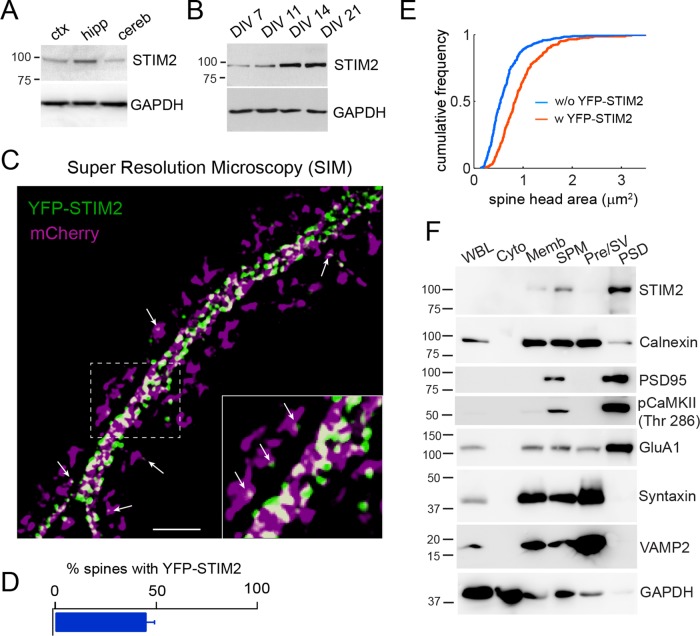
FIGURE 1:
STIM2 localizes to large dendritic spines. (A) Immunoblots of STIM2 from adult rat cortex (ctx), hippocampus (hipp), and cerebellum (cereb). (B) Developmental expression pattern of STIM2 in dissociated hippocampal neurons. (C) Superresolution microscopy (SIM) of hippocampal neurons (DIV 21) cotransfected with YFP-STIM2 (green) and mCherry (magenta). Scale bar, 5 μm. Arrows point to YFP-STIM2 puncta inside spine heads. Note that spine necks connecting spine heads to the dendritic shaft are not always visible on these high-resolution images. (D) Percentage of spines containing at least one STIM2 punctum. n = 711 spines from two independent experiments. (E) Cumulative distribution of spine size (area) with (n = 282 spines) or without (n = 415 spines) YFP-STIM2. (F) Fractionation and immunoblot analysis of adult rat brains. Equal amounts of protein were loaded in each lane. Cyto, cytosol; Memb, total membranes; Pre/SV, presynaptic membranes and synaptic vesicles; PSD, postsynaptic density; SPM, synaptosomes; WBL, whole-brain lysate.

