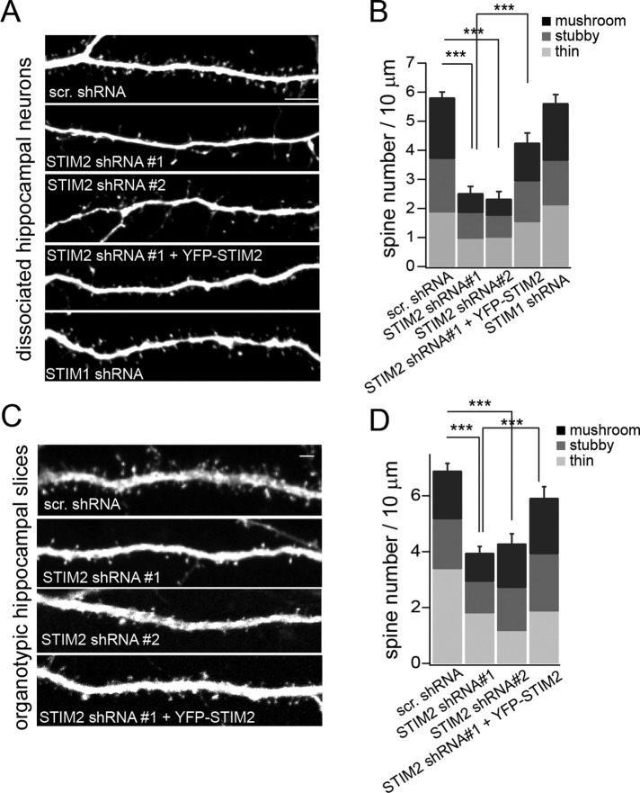
FIGURE 2:
STIM2 regulates spinogenesis. Spine analysis in dissociated hippocampal neurons (A, B) or hippocampal organotypic slices (C, D). (A) Confocal images of hippocampal neurons (DIV 21) coexpressing mCherry and the indicated shRNAs. For rescue experiments, STIM2 shRNA#1 was coexpressed with YFP-STIM2. (B) Quantification of spine density and spine type for conditions shown in A, using NeuronStudio software. At least 50 dendritic segments comprising >850 spines from three independent experiments were scored for each condition. (C) CA1 neurons were biolistically transfected with the indicated shRNAs or the STIM2 shRNA#1 together with YFP-STIM2 for rescue experiments. Spines were imaged in distal apical primary and secondary dendrites (see also Supplemental Figure S2). (D) Quantification of spine size and type. At least 45 dendritic segments comprising >1000 spines in three independent experiments were analyzed for each condition. STIM2 silencing decreased spine density in both dissociated cultures and slices. ***p < 0.001, ANOVA. The percentages of thin, stubby, and mushroom spines were not significantly affected by STIM2 shRNA#1 or shRNA#2; p > 0.05, ANOVA. Scale bar, 5 μm. See also Supplemental Figures S1 and S2.

