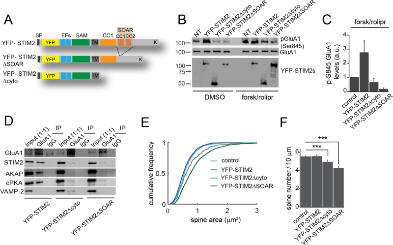
FIGURE 5:
The STIM2 SOAR domain mediates phosphorylation of and interaction with GluA1. (A) Cartoon showing the primary sequence of YFP-STIM2 WT, ΔSOAR, and Δcyto. (B–D) Cortical neurons (DIV 21) transduced with YFP-STIM2 WT and mutants. (B) Immunoblot analysis of GluA1 pSer-845 in cells expressing the indicated constructs and treated with DMSO (vehicle) or forsk/rolipr for 30 min. NT, nontransduced cells. (C) Densitometry analysis of pSer-845 after forsk/rolipr treatment from four independent experiments. (D) IPs from cells overexpressing YFP-STIM2 WT, Δcyto, or ΔSOAR with GluA1 Ab or control IgG. Lysates are the same used in B under DMSO condition. Note the marked decrease in endogenous STIM2, AKAP, and cPKA pulled down from cells expressing the STIM2 mutants. (E, F) Distribution of spine size (area) (E) and spine density (F) scored in neurons transduced with the indicated constructs or mock transduced. More than 400 spines from three independent experiments were analyzed for each condition. ***p < 0.001, ANOVA. See also Supplemental Figure S5.

