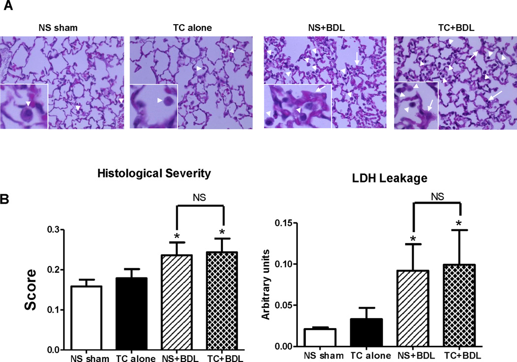
Figure 6. Lung injury in the surgical models.

(A) Representative HE sections at 40X from lung tissue obtained 24 hr after the surgical procedure. The insets zoom in on macrophages in the alveolar space (arrow heads) and alveolar septal wall thickening (arrows). (B) Overall histological severity (left) and lactate dehydrogenase (LDH) from bronchoavleolar lavage (BAL) fluid (right). (n=5 animals per group; lung tissue was graded over 20 fields at 40X). *, P<0.05, relative to the NS sham.
