FIGURE 1.

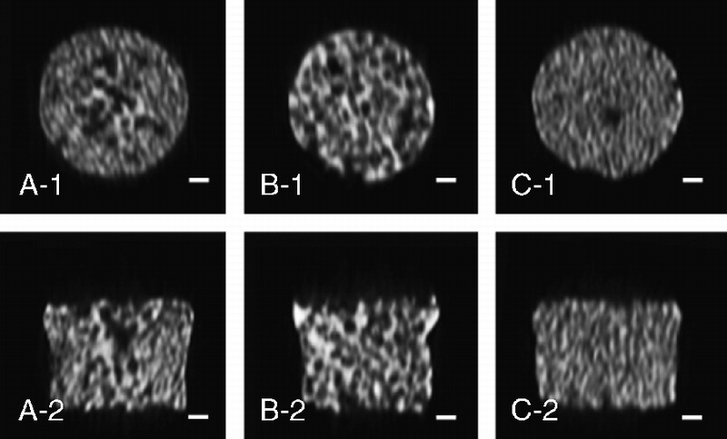
μCT images. A to C show images of the graded porous scaffolds (GP), the large pore scaffolds (LP), and the small pore scaffolds (SP). Scale bars = 1000 μm.

μCT images. A to C show images of the graded porous scaffolds (GP), the large pore scaffolds (LP), and the small pore scaffolds (SP). Scale bars = 1000 μm.