
Figure 2. Cleaved caspase-3 IHC in L-arginine induced pancreatitis.
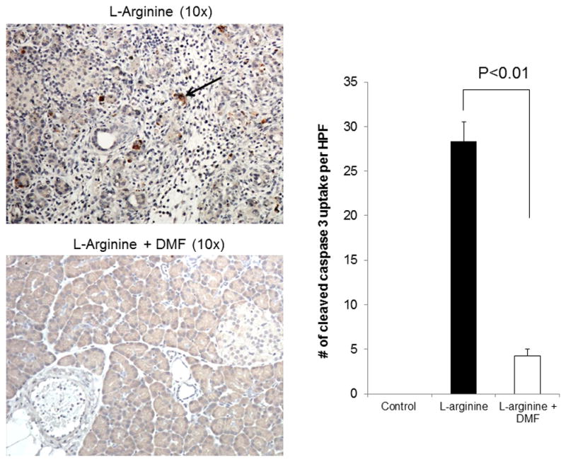
Representative photomicrograph of pancreas histology stained with cleaved caspase 3 antibody. In rats treated with DMF, the caspase-3 staining of the pancreas revealed significantly lower cleaved caspase 3 positive cells when compared with the L-arginine group (p <0.001). The arrow reveals an area of brown cytoplasmic uptake indicating cleaved caspase 3. The slides were reviewed by two blinded pathologists who scored the slides based on standardized criteria. Data are representative of three independent experiments.