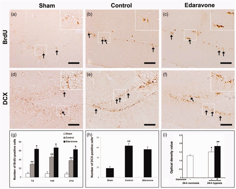
Figure 2.
Changes in BrdU- or DCX-positive cells in the SGZ and BrdU incorporation. (a) BrdU-immunoreactive cells in the SGZ of sham-operated group 7 days after sham-operation. (b, c) Representative images of the BrdU-immunoreactive cells in the SGZ 7 days after ischemia. BrdU-positive cells show brown granule. (d) DCX-immunoreactive cells in the SGZ of sham-operated group 7 days after sham-operation. (e, f) Representative images of the DCX-immunoreactive cells in the SGZ 7 days after ischemia. (g) Quantification of BrdU-positive cells in the SGZ. (h) Quantification of DCX-positive cells in the SGZ 7 days after ischemia. Arrows indicated positive cells. (i) Quantification of BrdU incorporation by spectrophotometry under different oxygen condition. The experiment was repeated three times. BrdU = 5-bromo-2-deoxyuridine; DCX = doublecortin. Scale bar = 100 µm. * p < .05, ** p < .01 compared with the control group. *p < .05. **p < .01 compared with the control group. #p < .05. ##p < .01 compared with the sham-operated group or normoxia. n = 5 in each group.

