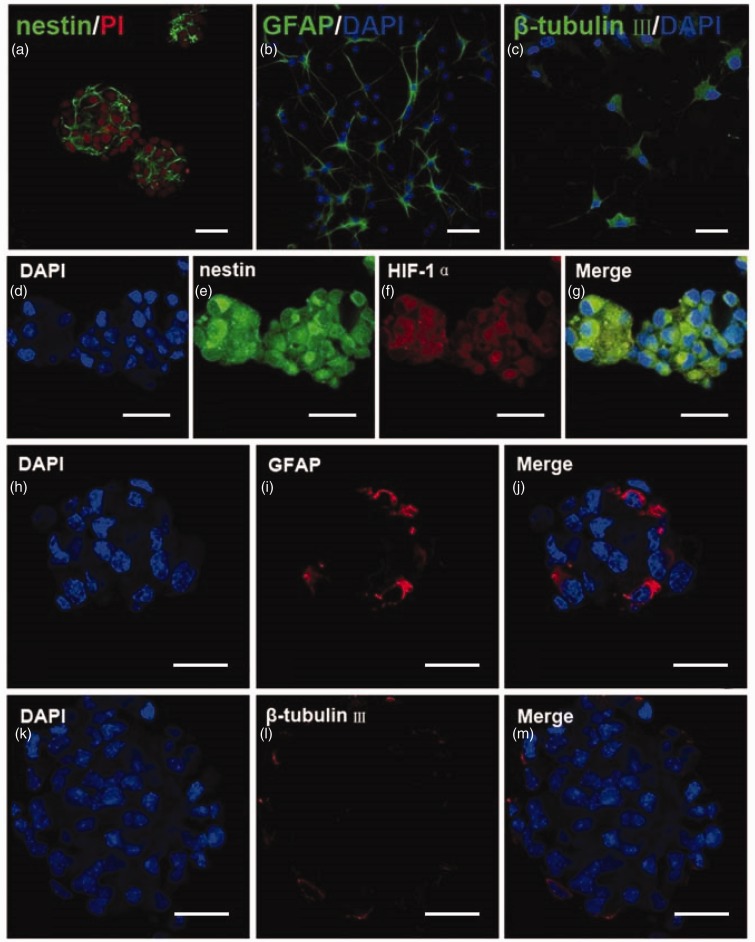
Figure 7.
Isolated cells were neural stem cells and passage 1 neurospheres retained the character of NSPCs after hypoxia. (a) Neurospheres formed after 3 days in culture and the vast majority of the neurospheres were nestin-positive (green). (b, c) A subpopulation of NSPCs expressed GFAP (green) or β-tubulin III (green). The nuclei were counterstained with PI (a, red) or DAPI (b, c, blue). Bar = 50 µm in panel (a) and (b). Bar = 30 µm in panel (c). (d–g) Confocal images of the passage 1 neurospheres double-labeled for nestin (green) and HIF-1α (red). The nuclei were counterstained with DAPI (blue). Most of cells in the passage 1 neurospheres were nestin and HIF-1α immunoreactive after 24 hr of hypoxia. (h–j) Confocal microscopic images of the passage 1 neurospheres labeled for marker of astrocytes, GFAP (red). (k–m) Confocal microscopic images of the passage 1 neurospheres labeled for marker of neurons, β-tubulin III (red). The nuclei were counterstained with DAPI (blue). GFAP = glial fibrillary acidic protein; HIF-1α = hypoxia-inducible factor 1α Bar = 50 µm.

