Figure 7.
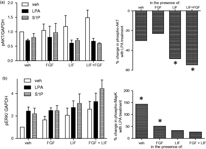
Costimulation with LIF and bFGF alter the effect of LPA and S1P on Erk and Akt signaling pathways.
hNP cells were grown in the absence of bFGF for 24 hr and were then stimulated with 20 ng/mL bFGF, 10 ng/mL LIF, 1 µM LPA, or 0.1 µM S1P as indicated. (a) To assess Erk Map kinase activation, cells were treated for 10 min and then harvested for Western blotting analysis with anti-phospho p42/44 Erk Map kinase antibodies. Expression of GAPDH protein was determined as a housekeeping standard. Band intensities were quantified and Erk phosphorylation levels were normalized to GAPDH levels. (b) To assess Akt activation, cells were treated for 30 min and then harvested for Western blotting analysis with anti-phospho serine 473 Akt kinase antibodies. Expression of GAPDH protein was determined as a housekeeping standard. Band intensities were quantified and Erk phosphorylation levels were normalized to GAPDH levels. Right panels: The ability of LPA to regulate Erk and Akt activation levels was compared when added to cells alone or in the presence of bFGF, LIF, or LIF and bFGF. Under each condition, the percent change in phosphorylation in LPA-treated cells versus vehicle controls was determined. *p < .05.

